Showing posts with label Rehabilitation. Show all posts

Wednesday, December 3, 2014

Low Back Pain: Rest vs. Exercise

Dear friend,

in today's blog I will address a problem that approximately 80% of Americans are faced with at least once during their lifetime and that many live with day in day out: Low Back Pain (LBP) aka lumbago.

This blog will provide some insights into the most common causes and treatment options of LBP and investigate whether rest or activity is more helpful when dealing with acute or chronic back pain.


The most common causes in younger adults:
  • Injury or overuse
    A sudden onset of pain and stiffness often after activity, a sudden movement or heavy lifting, is mostly caused by soft tissue injuries to muscles and ligaments in the back and along the spine such as muscle strains or sprains. Other causes that fall into the category of injury or overuse are injuries to the small facet joints of the spine or fractures of the bony processes of the spine (attachment points for muscles and tendons). Both types of injuries commonly heal on their own. A combination of rest, heat/ice applications, anti-inflammatory drugs and low back exercises improving spinal stability can be helpful treatment options.
  • Nerve root compression
    An Acute or chronic nerve root compression can be caused by a variety of problems. Some of the more common causes in younger adults are:
  • A herniated disc: As discs degenerate and break down, the inner core of the disc starts to leak out through the outer portion applying sudden pressure on nerve endings.
  • Isthmic Spondylolethesis occurs when a vertebra slips forward causing instability and nerve root compression. The reason for the slippage is often a small fracture in a piece of bone that connects the two joints on the back side of the spinal segment. Th fracture typically happens during childhood, starting to cause trouble in young adulthood.
  • Lumbar Degenerative Disc Disease: Even though this is a degenerative disease it often affects young adults in their early 20's. It refers to the breakdown of discs in between vertebrae leading to instability and inflammation.
  • Sciatica: All of the previously mentioned causes of nerve root compression if effecting any of the major lumbar nerves can lead to Sciatica . Sciatica refers to radiating pain, weakness and numbness along the pathway of the nerves into the buttocks, legs and even feet. The pain can be worse in the feet and legs than in the back and is certainly more intense than the often dull aching pain resulting from strains or sprains.


    The most common causes in older adults:
    While older adults can experience the same pain related to the problems that apply to younger adults, they are more likely to experience chronic pain related to degenerative processes of the spine and it's joints.


    Degenerative Processes

    • Osteoarthritis effects most commonly the small joints of the spine, like the facet joints and the Sacro-Iliac joint between sacrum and ilium at the bottom of the spine. These degenerative joint changes (wear and tear on protective cartilage layers) can lead to inflammation of the joint area itself, causing instability, pain, and stiffness. In addition it can lead to the growth of bone spurs which again can apply pressure to the nerve roots. Osteoarthritis commonly effects other joints in the body as well, such as hips or knees causing gait changes leading to secondary pain in the low back due to compromised body mechanics.
    • Spinal Stenosis refers to the degeneration of discs, vertebrae, muscles and tendons that make up the spinal column. This degenerative processes can lead to a choking (greek=stenosis) of the lumbar nerves causing increased leg pain with walking (pseudoclaudication) which improves almost immediately when sitting down.
       
    • Osteoporosis is a condition in which the bone density degrades to the point of bone fractures. This condition is most common amongst post menopausal women. One of the most effected areas is the lumbar spine. Osteoporosis in the lumbar spine can result in compression fractures of the vertebra causing significant low back pain.
    Less commonly found causes for low back pain worth mentioning are Ankylosing Spondylitis, bacterial infections and tumors of the spine.


    Treatment Options:
    • Heat and Cold applications are usually the first line of defense patients with low back pain are reaching for. Cold packs and ice have been hailed for many years as anti-inflammatory (antiphlogistic) and pain reducing (analgesic) treatment options. It is recommended to apply cold/ice for 15-20 minutes for the first 48 hours after injury. A newer line of thought questions the value of ice or cold packs as a form of anti-inflammatory treatment, because it leads to reduced blood circulation in the inflamed area, reducing the number of white blood cells (the body's natural defense against inflammation) at the source of inflammation. However the cold will numb nerve endings and reduce impulse transmission speed validating it's analgesic qualities. Try a damp cloth between skin and cold source for better conductivity and better results. If introducing ice as an anti inflammatory treatment I would recommend short ice massages. (Ice massage for LBP relief)  The short introduction of cold triggers greater blood flow to the area and lead to an increase in the number of inflammation fighting white blood cells.

      Heat applications are indicated if the source of discomfort stems primarily from muscle spasms. The heat will relax the muscles and promote healing. Don't use heat on injured areas that seem swollen and/or red.

      If circulatory issues or nerve damage are present do not use either form of treatment. Even though cold/ice is often recommended for the first 48 hours I suggest you try both, heat and ice and find out which provides greater relief.
    • Topical lotions/cremes/rubs are in most cases not much more than a distraction from the pain as they commonly promote surface heat rather than penetrating heat. Those containing Capsaiscin, an extract from red chilli peppers, do effect nerve impulse transmission and can be a helpful addition to analgesic medicine in treating pain. Capsaiscin's effect is cumulative and therefore requires a prolonged period of usage before its full benefit is noticeable.
    • Pain medications (Analgesics) reach from over the counter pills like Tylenol to powerful skin patches prescribed one at a time. In severe chronic back pain cases these types of analgesics are often combined with codein causing possibly addiction. Antiphlogistica include non-steroidal anti inflammatory drugs (NSAID) such as Advil, Aleve or Aspirin. NSAID address pain and inflammation. The last group of medications commonly administered for acute and chronic back pain are muscle relaxants. Muscle spasms appear quite often as a result of nerve root compression. Spasms add to pain, reduce circulation and slow the healing process. Muscle relaxants may cause drowsiness and should preferably be taken at bed time.
    • Invasive Treatment Methods
      This form of treatment includes most often epidural injections; commonly a powerful mixture of an anti-inflammatory steroid (cortisone) and a local anesthetic used to calm nerve root irritation and reduce swelling.
    • Physical Therapy
      In case of an acute injury such as a fracture or a herniated disc the RICE injury protocol as is typically the best choice. Rest, Ice, Compression and Elevation are indicated to prevent further damage/injury and manage irritation and inflammation. This protocol is most effective during the first 48-72 hours and typically followed up with passive Physical Therapy modalities such as cold/heat (see above), Iontophoresis, trans-cutaneous electrical nerve stimulation (TENS), and ultra sound.

      Iontophoresis is a modality in which steroids are delivered through the skin using an electrical current. A TENS unit is used to override painful nerve impulses to the brain reducing pain. Ultra sound is a form of deep heating soft tissue often successful in reducing acute pain and enhancing soft tissue healing.

      Education and Exercise
      A trained Physical Therapist, Physio-Therapist or Kinesiologist can help identify poor body mechanics or postural deviations, which may be contributing factors to your LBP. Together with the therapist you will develop learn to implement strategies that can help to improve those movement and/or postural issues. Learning about proper lifting techniques, a proper work station set-up, and postural awareness can positively influence the outcome of rehabilitation and secondary prevention.

      Postural deviations and poor body mechanics are often caused by a combination of a lack of muscular strength and shortened muscles leading to reduced range of motion in the large joints. Poor range of motion especially in the lumbar-pelvic-hip region of the body places undue stress on the spine, as it often requires the spine to compensate for immobility in hips and the sacroiliac joint. The combination of poor mobility and a significant lack of muscular strength leaves the spine vulnerable to injury and increased wear and tear. Physical Therapy will address these issues by leading you in an exercise program that will improve spinal stability and stretching exercises that will increase functional range of motion.
    • Examples:
      Supine Floor Bridge: Lie on your back with your knees bend at a 90 degree angle and soles of feet firmly placed on the floor. Tighten your stomach and buttocks. Slowly raise your pelvis off the floor as far as you can, maintaining neutral spine throughout the movement. Hold at top for specified time and slowly return to floor. Careful if you have neck issues.


      Body Plank Prone
      : Lie down flat on your stomach. Tuck your toes under and place elbows next to your shoulders. Lift body off the floor supporting plank body position on toes and elbows. Keep stomach and buttocks tight to avoid sway back. Hold for specified time and slowly return to floor.


      Clam Shell Exercise
      : Lie down on your side, placing shoulder over shoulder, hip over hip, knee over knee, and ankle over ankle. Split off top knee keeping feet together and hip over hip (clam shell movement).


      Dead Bug Exercise
      : Lie down on your back, secure spine by contracting abdominals and glutes. Bend both hips and knees at 90 degrees and reach arms straight up towards ceiling. Lower right arm and left leg maintaining neutral spine. Return to starting position and use other diagonal.


      Hamstring Stretch
      : Lie on your back, both knees bent, feet on the floor. Straighten one leg out until you feel stretch on the back side of the thigh. Hold stretch for prescribed time and repeat with other leg.


      Kneeling Hip Flexor and Quadricep Stretch
      : Kneel down on one knee placing other foot in front. Tighten abdominals and glutes and push pelvis straight forward shifting body weight to front foot. You should feel stretch in front of hip and thigh. Hold stretch for prescribed time and repeat with opposite leg.


      Please consult your physician before starting any exercise program. Keep in mind that any exercise program should be designed with individual needs in mind and be instructed and supervised by a trained health professional.
    • Rest or Exercise?
      Over the past four decades the rehabilitation protocol for LBP has clearly shifted from rest, sometimes total bed rest, to a more active path to recovery. Despite this change in approach the actual course of action is still determined by the cause of your back pain. Exercises can be beneficial and recommended, or strictly contraindicated if exercising carries a high risk for further damage. Generally the first course of action is to rest and have a physician assess the cause of your back pain and develop a treatment plan. Acute injuries such as compression fractures of the vertebra or spinous processes as well as a herniated disc require immediate rest to avoid further injury and possible nerve damage. Other problems such as strains and sprains of muscles and ligaments in the low back and along the spine often respond positive to gentle forms of stretching and low impact aerobic activities as both can be helpful in reducing muscle spasm. Regular exercises are of great importance in managing chronic back pain often caused by degenerative changes of the spine. Chronic back pain does require an individualized exercise program to stabilize and mobilize the spine as well as the lumbar-pelvic-hip region in order to reduce pain and improve function and movement. Secondary back pain is often the result of postural and gait deviations caused by osteoarthritis to other large joints such as knees and hips. In order to reduce the back pain an exercise program needs to manage the original cause first and later correct gait and posture.
    Conclusion:
    LBP is responding in most cases positive to exercising and often can be a valid alternative to surgical intervention. The starting point, the intensity, the progression as well as the choice of exercises should be determined by a team of Health Professionals experienced in the rehabilitation and management of LBP.

    I hope this article is providing you with some helpful information in dealing with your own low back pain or that of a loved one. As always, your questions and comments are greatly appreciated.

    A Sante,

    Hartmut 


    Sources:
    http://www.nytimes.com/health/guides/symptoms/back-pain-low/exercise-and-physical-therapy.html
    http://www.injurytreatment.com.au/search-injury-information/back-spine/lumbar-spine
    http://www.webmd.com/back-pain/features/relieve-back-pain-with-core-strength-training
    http://www.spine-health.com/wellness/exercise/back-strengthening-exercises
    http://www.bigbackpain.com/back_pain_treatment.html
    http://www.webmd.com/back-pain/tc/low-back-pain-prevention
    http://www.webmd.com/back-pain/tc/low-back-pain-cause
    http://www.ncbi.nlm.nih.gov/pmc/articles/PMC3809228/
    https://www.inkling.com/read/acsms-guidelines-exercise-testing-prescription-9th/chapter-8/low-back-pain
    http://www.spine-health.com/blog/bed-rest-or-staying-active-better-low-back-pain-recovery
    http://www.spine-health.com/treatment/physical-therapy/physical-therapy-low-back-pain-relief

    Sunday, August 25, 2013

    Osteoarthritis of the shoulder


    Dear Reader,

    Like my mother many people suffer from chronic severe shoulder pain caused by degenerative joint disease also known as osteoarthritis.

    Osteoarthritis occurs when the bone protective- or articular cartilage wears down and causes swelling, inflammation, and pain. In some cases bone spur (osteophytes) growth might occur, as bone rubs against bone. The shoulder is made up of two joints, the acromioclavicular (AC) joint and the glenohumeral joint. The AC joint is located where the clavicle (collarbone) meets the acromion at the tip of the shoulder blade. The glenohumeral joint is the meeting point between the arm bone (humerus) and the scapular (shoulder blade).

    In the majority of cases osteoarthritis of the shoulder or any other joint for that matter is found in people age 50 or older. In the earlier years osteoarthritis is typically caused by a traumatic injury to the joint. A very small percentage of young osteoarthritis patients have a genetic predisposition to the joint disease that causes an early onset.

    When seeing an orthopedic specialist, he/she will most commonly start the diagnostic process by establishing a medical history and perform a physical exam looking for swelling, pain and tenderness. Often muscular weaknesses surrounding the joint is found due to pain induced inactivity. In addition x-ray and MRI can produce imagery that details the degree of degeneration of the joint more accurately.

    Osteoarthritis of the shoulder can be treated conventionally, through medication, and surgically. The first step of treatment is to rest the shoulder and modify activities involving the shoulder. The application of ice or heat packs to reduce swelling, inflammation and pain and improve mobility are typically temporarily and do not eliminate the cause. Your physician might prescribe a physical therapy program focused on  stretching and strengthening muscles to improve joint range of motion.

    Should these conventional methods fail to bring relief medications such as Non-Steroidal-Anti Inflammatory-Drugs (NSAID's: Ibuprofen, Naproxen, Aspirin) or Cox2 Inhibitors (Celebrex) can  be used to further reduce inflammation, swelling and pain. Advanced stages and severe shoulder pain is often treated with corticosteroid or hyaluronic acid injections. Steroid injections reduce swelling and thereby alleviate pain and decrease joint stiffness. Hyaluronic acid injections are known as "viscosupplementation" and help lubricate the joint similar to the way W40 works on mechanic joints.

    If all non-surgical options are exhausted your physician will suggest either a total shoulder replacement (arthroplasty) or a partial replacement in which only the head of the humerus is replaced (hemiarthroplasty). Both these surgeries are performed to treat osteoarthritis of the Glenohumeral joint. The removal of a small section of the end of the collarbone known as resection arthroplasty is the common surgical choice to treat osteoarthritis of the AC joint.

    In recent years stem cell research has created new hope for osteoarthritis patients. In the last years stem cells who help heal and regrow cartilage were used in the repair of small cartilage defects. Currently studies are ongoing to explore whether the same treatment can help grow whole joint surfaces such as knees and hips in a lab to resurface the degenerated joint. For now this is still science fiction but there is a great deal of optimism and reason to be hopeful.

    Many of my arthritic clients ask whether exercising is beneficial and whether I would recommend exercising while in pain. My answer to both questions is YES.

    Our joints need movement! Movement nourishes and promotes joint health. Maintaining strong muscles will help support the weakened joint structure and reduce joint stress as you move. Stretching muscles and utilizing gentle mobilization exercises will help maintain good functional range of motion allowing for optimal use of the joint. Cardio-vascular exercises help in the prevention of diseases commonly known to be associated with inactivity and manage body weight therefore reducing impact on spine and joints. In addition learning meditation and relaxation techniques have shown to reduce stress and pain.
    The exercise choices and the workloads when exercising with osteoarthritis depend on the severity of the disease as well a the timing. Workloads and work-out intensities have to be reduced when training an arthritic joint. Certain exercises might require modifications or need to be avoided all together in order to reduce the risk of causing aggravation. During an acute phase of inflammation one should further reduce or even eliminate the workload and focus instead on flexibility and mobility exercises working around the pain rather than through the pain.

    Exercise suggestions for Shoulder OA:

    Overhead Shoulder Stretch:
    Stand with feet shoulder wide apart, arms hanging in front of body. Interlock fingers with palms facing down. Stretch arms straight overhead with fingers interlocked and palms facing ceiling.

    Hold stretch for 20-30 seconds
    2-3 Repetitions


    Crossover Shoulder Stretch:
    Stand with feet shoulder wide apart. Reach with one arm across opposite shoulder bending elbow. Gently push elbow further towards opposite shoulder.

    Hold stretch for 20-30 seconds
    Repeat 2-3 times each side


    Towel Shoulder Stretch
    Feet shoulder wide apart. Hold towel in one hand, drop it down the spine, Grab low end with other hand and gently pull top hand down behind head.

    Hold stretch 20-30 seconds
    Repeat 2-3 times each side


    Shoulder Blade Pinch:


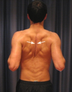
    Stand with feet shoulder wide apart. Draw shoulders down and back until you feel your shoulder blades pinch in the back.

    Hold for 5-10 seconds
    Repeat 5-10 times


    Rotator Cuff Strengthening
    External Rotation

    Stand with feet shoulder wide apart. Arms bend by your side at 90 degree angle. Grab elastic band with one hand and rotate outward. .

    Repeat 10-20 times each side


    Internal Rotation


    Same position as before but this time arm rotates inward

    Repeat 10-20 times each side.


    These exercise are only a small selection and might not be suitable for everyone. Please talk to your physician and/or physical therapist before starting any exercise program.

    I hope this short article is helping you understand your own condition or that of someone close to you. If you have any comments or questions please don't hesitate to email me.

    Best,
    Hartmut


    References:


    Web Md: http://www.webmd.com/osteoarthritis/guide/shoulder-osteoarthritis-degenerative-arthritis-shoulder
    American Academy of Family Physicians: http://www.aafp.org/afp/2008/0901/p605.html
    About.com: http://osteoarthritis.about.com/od/shoulderosteoarthritis/a/shoulder_OA.htm
    Arthritis Health: http://www.arthritis-health.com/types/osteoarthritis/shoulder-osteoarthritis-treatment
    NCBI: http://www.ncbi.nlm.nih.gov/pubmed/23190869
    Science Daily: http://www.sciencedaily.com/releases/2013/01/130124163246.htm
    Chicago Tribune: http://articles.chicagotribune.com/2013-04-24/health/sc-health-0424-arthritis-stem-cells-20130424_1_stem-cells-farshid-guilak-cartilage
    Arthritis Foundation: http://www.arthritis.org/conditions-treatments/disease-center/osteoarthritis/

    Wednesday, January 16, 2013

    Warm-Up and Cool -Down: Why and How

    Dear Friend,

    I wish you and your family a Healthy and Happy 2013.

    Yesterday I watched my son's High School soccer game. As so often, my observations triggered a new blog. During the middle of the game one of my son's teammates, who just a few minutes earlier had entered the game as a sub, went down on to the pitch in a great deal of pain. No one knew exactly why. I thought it was the lack of proper warm-up before he entered the game that caused the injury. Turns out I was wrong. Upon further examination the boy showed all classical symptoms of Sciatica. However having observed his warm-up prior to coming into the game made me want to write a blog discussing the changes that have taken place over the past 4-5 years with respect to warm-up and cool-down and the scientific reasoning behind these changes.

    The right warm-up and a proper cool-down need to be essential parts of every work-out and athletic competition. Correctly done they help minimize the risk for injury, increase physical performance and help with recovery after the competition or work-out.

    The preferred warm-up of the past and that of my son's teammate included some form of gradually increased cardio vascular activity ( in his case way too little) like running, cycling, swimming etc. to increase the body's core temperature and blood circulation within the muscles. This phase was followed by static stretches to increase flexibility and range of motion (prolonged stretches that are used to increase the length of soft tissue and flexibility of the muscle). This warm-up procedure is flawed in at least two ways.
    1. Static stretching is a very inactive warm-up method which leads to a decrease in muscle and body-core temperature that had just been elevated during the first phase of the warm-up. Therefore rendering the cardio-vascular warm-up part worthless. Reducing muscle temperature and core-temperature reduces physical performance ability (reduced oxygen supply, reduced neurological activation level) and increases risk of soft tissue injury as a colder muscle is less pliable/flexible and tears easier.
    2. Besides that, several recent studies have shown that static stretches reduce the muscles ability to contract forcefully leaving that stretched out muscle weakened for up to an hour. This muscle weakness leads to an increased risk for injury and a muscle performance reduction.
    Does this mean static stretches should be avoided all together? The answer is no. Static stretches are still a vital part of an athletes overall training. They do increase flexibility and improve joint biomechanics thereby reducing risk for injury to soft tissue and joints. Static stretches should based on today's knowledge be performed as part of a cool-down procedure and/or be done on off days.

    Lately we are leaning more and more towards sports specific dynamic warm-up programs. These dynamic warm-ups focus more on the neuromuscular systems of the muscle. Dynamic warm-ups help with short term flexibility gains and lead to more adequate protective reflex responses from the Golgi Tendon Organs without compromising the strength of the muscle. Golgi Tendon Organs are lying deep within the muscles and protect the muscles from being overstretched. If not properly prepared and activated during warm-up these organs often over react during sports activities leading to muscle strains. Dynamic warm-up protocols help dampen the reaction of the Golgi Tendon Organs leading to improved muscle performance.

    To properly warm-up I suggest you continue to start the same way you used to. Brisk walking, jogging or cycling etc. will help increase core and muscle temperature, improve oxygenation of muscle, and get your metabolic and nervous systems ready for action. After that skip the static stretches and move into dynamic sports specific exercises such as high knee jogging, butt kickers, multi directional lunges, bear walks, leg swings, arm circles etc.

    After completing your work-out or competition slowly turn the system back down by gradually decreasing the intensity. Slow jogging and static stretches are a good way of returning physically and mentally to a real life activity level.

    I hope I was able to shed some light on the recent changes to Warm-Up and Performance Readiness. As always, if you have any questions or comments please feel free to contact me.

    A Sante,
    Hartmut


    Sources:
    Mayo Clinic: http://www.mayoclinic.com/health/exercise/SM00067
    Journal of Strength and Conditioning Research. ” 2008:
    http://hpcsport.com/publications/staticstretching2.pdf
    About.com: http://sportsmedicine.about.com/cs/injuryprevention/a/aa071001a.htm
    IDEA: http://www.ideafit.com/fitness-library/pre-run-stretch-hampers-endurance

    Sunday, June 17, 2012

    IT Band Syndrome

    Dear Friends,

    today I want to address a very common and painful condition effecting the outside of thighs, hips and knees. The Iliotibial Band (IT Band) runs from the hip (ilia crest) along the outside of the thigh attaching to the tibia (shinbone) just below the knee. It stabilizes the knee and coordinates muscle activity while moving. This band can become irritated and inflamed causing pain at hip and knee.

    IT Band Syndrome (ITBS) is common in long distance runners, cyclist and tennis players caused most often by
    • overuse/over training
    • increasing workload/distance to soon
    • returning from an injury before healed
    • training errors
    • poor biomechanics or anatomical problems

    While the first three causes are pretty much self explanatory the last two might need elaboration. Training errors amongst runners can include running on the same side of the road or on the track in the same direction. By keeping same leg on the inside turn  you are causing excessive stress on the outside leg. Most roads are crowned, meaning the surface slopes to the outside edges causing a lateral pelvic tilt. Other biomechanic problems include super pronation, bow legs and significant leg length discrepancies.

    The cardinal symptom of ITBS is knee pain as the IT Band slides across the femoral condyle on the outside of the knee. Pain can extend along the thigh up to the hip. Pain typically is worse at heel strike while running and when ascending/descending on stairs, or getting up from seated position.

    Treatment of this condition initially involves Rest, Ice, Compression and Elevation (RICE) in combination with Physical Therapy, stretches  and mobilization of IT Band. In addition Non Steroidal Anti Inflammatory drugs might be prescribed or in severe cases Cortisone Injections given.

    Even though there are only insufficient randomized, controlled trials as to which particular exercises are most beneficial in reducing the IT Band Syndrome and prevent recurrence some studies amongst specific groups of patients (i.e. long distance runner) seem to indicate that stretching the IT Band, mobilization of the IT Band and single leg strengthening exercises have been successful in the secondary prevention of ITBS.

    Here are some recommended exercises I'd like to share with you:
    1. Standing ITB stretch
    2. Advanced ITB stretch (Pigeon Pose)
    3. Foam Roller ITB mobilization
    4. Hip Hike on step
    5. Single Leg Squat
    6. Side Lying Hip Abduction
    Since ITBS is most often caused by overuse cross training can be successful in reducing the risk of acquiring such injury.

    I hope this little bit of information was helpful and will keep your IT Band in great shape.

    A Sante,
    Hartmut

    References:
    About.com Sports Medicine
    emedicinehealth.com
    VHI's Evidence-Based Exercise Prescription Newsletter